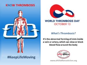
Today is World Thrombosis Day.

aHUS is the result of thrombosis in the small blood vessels – thrombotic microangiopathy, or TMA.
To mark World Thrombosis Day, ThrombosisUK, a charity for those affected by thrombosis held an international conference in London about “Thrombosis and Thromboprophylaxis in Pregnancy and the Puerperium”
The alliance attended the Conference and a synopsis of it follows.
The morning talks sessions were about the profiling of potential patients who were at higher risk of thrombosis, diagnosing someone with a thrombosis and providing effective treatment for a thrombosis.
Thrombosis in pregnancy is a leading cause of death but it is also a rare event
Although risk profiling was well established much more development was needed because more than half of potential thrombosis patients were not being picked up with existing risk criteria.
Similarly, the sensitivity and specifity of existing diagnosis decision rules is not precise enough for identifying the sort of thrombosis event happening.
The effectiveness of front line treatment warfarin, heparin (familiar to aHUS dialysis patients) varies between type of clotting and between those who are pregnant and those who are not. Also, the wearing of compression stockings has diminishing effect the longer it is prescribed after the intial event and the level of compliance with wearing them by the patients.
Within the clinical communities there was a considerable degree of consensus on guidelines on Thromboprophylaxis as a clinician from New Zealand, Dr Claire McLintock, illustrated this in her talk, but work was still needed to get better outcomes.
In the afternoon, the Conference split into two breakout groups; and the alliance joined the “TMAs in Pregnancy” group.
aHUS in pregnancy is one of the top issues in the aHUS community as the trigger of roughly one in ten aHUS patients is down to pregnancy.
The session was chaired by Dr Kate Barnham and Prof. Beverly Hunt. Prof. Hunt opening remark was that the session was very important because women were still dying from TMA in pregnancy through lack of diagnosis. She then illustrated the difficult challenge of diagnosis with the Campistol Group’s four overlapping circles diagram of the spectrum of TMAs.
The most common TMA in pregnancy is HELLP/PET in which pre-eclampsia the breaking of the placenta and endolethial disfunction is the mechanism but the reason it happens is not yet conclusive. As it is the most common TMA in pregancy it would be the first considered ; but as the presenter Dr Lucy Mackillop concluded there needed to be “flexibility in diagnosis” as other TMAs remain possible, and that TTP particularly should be thought about. It tends to occur later in pregnancy and with pre-eclampsia “delivery” is the treatment of choice, and after it the mothers recover.
Prof Marie Scully, who is member of the Scientific Advisory Board of the aHUS Registry, then talked about TTP, a TMA caused by ADAMTS13 deficiency, and which is triggered by pregnancy in 10% of all TTP incidents. It tends to happen in the late second and third trimester of pregnancy and plasma exchange can be an effective treatment. Due to success in managing women with TTP through pregnancy, termination is no longer advocated but patient counselling is important.
Antiphospholipid Syndrome (APS) mediated TMA is caused by an acquired autoimmune response and it has a varied spectrum of disease manifestations. Speaker Dr Karen Breen revealed that an acquired immune auto antibody exists in 5% of the general population, but appears at much higher levels in those affected by APS. It is increasingly thought that Complement activation is implicated with recent research revealing higher levels of C3a and C4a, an inflammation response, with higher complement tick over and some use of eculizumab is being tried. No specific bio-markers exist and it was a case of the past is the best predictor of the future. APS happens though out the pregnancy period resulting in miscarriages and premature delivery.
Prof. Liz Lightstone then talked about TMA caused by Lupus and Vasculitis, although because the latter was not seen as a cause of TMA she focused on Lupus. The symptoms of Lupus onset would be very familiar to those who have experienced aHUS. A kidney biopsy would be the important way to diagnose Lupus but a biopsy is not encouraged in pregnant women. The usual question would be “is this pre-eclampsia?” not “Is this TMA?” Lupus with TMA meant the worst possible outcomes. Schitsocytes on a blood film would distinguish TMA from a Lupus flare. Like APS there is some thought that Complement is playing a role and some use of eculizumab is being tried.
In a talk about aHUS diagnosis and management in pregnancy, nephrologist’s Dr Kate Bramham said the kidney gets bigger in pregnancy to create more creatinine clearance and questioned whether there is more non- eclampsia TMA than currently thought. aHUS in pregnancy is a differentiated diagnosis, it is what is left after all else is discounted, and after all it only happens in 1 in 25000 pregnancies. Complement is activated during pregnancy which is natural and not a problem for those who do not have a pre- disposition to unregulated complement activity.
Having set the scene Dr Bramham handed over to Prof David Kavanagh from the National Renal Complement Therapy Centre in Newcastle upon Tyne. He briefly explained the genetics of Complement and the amplification loop of the alternative pathway that leads to the activation of the Membrane Attack Complex which goes uncontrolled in aHUS because of mutations in the Complement control components. He reported on the results of studies from aHUS patient databases in France/UK/Italy and Spain from which 16% of aHUS patients presented because of pregnancy and 76% of these were post-partum.
After a break for tea, this was the UK after all, a discussion took place on whether an algorithm could be developed to give guidelines to a TMA diagnosis in pregnancy. Starting with a TMA diagnosis decision chart for the whole TMA cohort the experts talked about what specific questions and tests results would need to be asked at the outset to distinguish between the types of TMA in pregnancy covered in the conference. This was not an easy thing to do but an initial stab at it was made and will be taken away for development.
As an afterthought risk profiling although not foolproof can lead to thinking TMA earlier, similarly trimester/ post partum timing of onset can guide some differentiated diagnosis. Then it is down to the blood and other tests.
There will be videos of the conference talks eventually on the ThrombosisUK website.
In the meantime, the aHUS alliance video of the talk given by Dr Craig Gordon “Pregancy and TMA- a challenging dilemma” at it s TMA symposium in the Harvard Medical School Conference Centre in August can be seen at this link here.
Also, the discussion between Prof. Fadi Fakouri and aHUS patient Megan Berry about pregnancy aHUS issues can be read at this link here.
With all the information and knowledge that has become available in the past few years it begins to beg the question about the status of research in answering the question “Do aHUS families have all the correct information to make informed family planning decisions?”


